After several years of data collection and analysis, a team from IGBMC has just published its research on the study of eukaryotic ribosomes in complexes with 16 molecules that inhibit their activity. Ribosomes are essential for cell survival as they are the "micro-factories" that perform protein synthesis by reading the information encoded in genes. Until very recently, our understanding of ribosome inhibitors in eukaryotes was very limited. These results, which have been published in the journal Nature, are crucial to foresee the development of new drugs against infectious diseases, cancer and genetic disorders.
While some proteins have a structural role (such as collagen, keratin, and actin), others are antibodies, hormones (insulin, pituitary hormones) or even enzymes. They are responsible for almost all the functions of both bacterial and eukaryotic cells, and of the body as a whole. All things considered, it comes as no surprise that ribosomes, those complex molecular complexes responsible for the protein synthesis, are under close scrutiny. The malfunctioning of protein synthesis can indeed have a tremendous impact on the cell, and is involved in diseases such as cancer and certain genetic disorders. On the other hand, blocked synthesis quickly results in cell death, which explains the use of ribosome-targeting antibiotics in bacteria (half of current antibiotics target bacterial ribosomes). This is one of the reasons why the structural information available relates to ribosomes in bacterial species. Another reason is that eukaryotic ribosomes are at least 40% larger, which further increases their complexity and makes them even more difficult to study.
IGBMC has been focusing its research on the well-known yeast Saccharomyces cerevisiae for several years, and published its results in the prestigious journal Nature in September 2014.
The researchers had already obtained the structure of "isolated" ribosomes, and with these new results, they have now unraveled the structure of ribosomes in successive complexes with 16 different molecules. Crystal diffraction images of ribosome/inhibitor complexes were collected on SOLEIL's PROXIMA 1 beamline, which is itself quite the technological achievement, since very few beamlines in the world allow researchers to determine the structure of macromolecular complexes as large as a ribosome, and have high enough resolutions to identify very specific areas composed of but a few atoms-in this case, the binding sites of inhibitors.
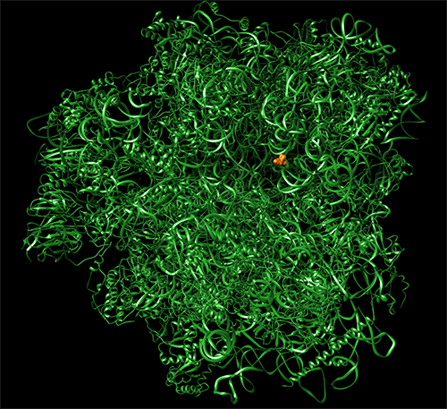
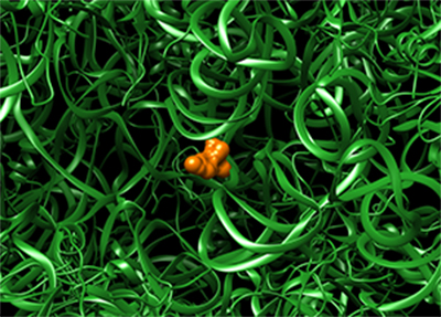
Figure caption: 3D structure of the complex between a S. cerevisiae ribosome 80S subunit (199360 atoms), in green, and a molecule of the anisomycin antibiotic (19 atoms), in red.
The researchers also decrypted the mechanism involving inhibitors during protein synthesis. Through kinetic experiments and accurate localization of the inhibitors on ribosomes, the team found out the specific stages in which the inhibitors were involved during mRNA translation into protein. It appears that these stages vary according to the inhibitors' size (for a given family of inhibitors). This information could certainly prove extremely useful for future drug design.
Lastly, a better understanding of the mode of action of these molecules sheds light on their potential difference in effectiveness between prokaryotic and eukaryotic ribosomes. Understanding such differences is crucial to avoid side effects with certain antibiotics that, at times, disrupt the functions of the eukaryotic cell to be cured.
Specifically targeted eukaryotic ribosome inhibitors are not only precious subjects for the study of protein synthesis in cells, but also for the development of new therapies against infectious diseases, cancer and genetic diseases.