Crystal structure of part of the type VI secretion system solved on PROXIMA-1
Among the multiple systems used by bacteria to survive and multiply, the T6SS- type VI secretion system is one of the main ones found in so-called Gram-negative bacteria, which include many species potentially pathogenic to humans (eg E. coli, P. aeruginosa). T6SS is a complex of more than 10 different proteins that enable these bacteria to target and inject toxins into their "prey" (prokaryotic or eukaryotic) cells after perforating their envelope. To accurately understand the molecular mechanisms that govern the secretory activity of this complex, researchers from the Pasteur Institute and the CNRS studied the structure of each of its elements, including experiments on PROXIMA-1 and PROXIMA-2A. Their results were published in Nature.
Gram-negative bacteria have double membranes (inner/outer) separated by a space known as the periplasm. The TSS6 secretion system is anchored in the bacterium like a syringe that crosses this double membrane such that the "piston" side is on the inside, in the cytoplasm, while the other end, containing the toxin, points outwards aimed at the target. This is why researchers also compare TSS6 to bacterial nano-crossbows shooting arrows at their targets. The transmembrane portion, which anchors the complex in the bacterium, consists of three proteins, TssL and TssM anchored in the inner membrane, and TssJ, a lipoprotein, located in the outer membrane.
In order to know the precise structure of the different components of this complex, the research groups led by C. Cambillau, E. Cascales and R. Fronzes conducted biocrystallography experiments on the PROXIMA-1 beamline and PROXIMA-2A Beamline at SOLEIL. Part of the TssM protein structure was resolved on PROXIMA-1.
Transmembrane proteins are difficult to crystallize; the researchers worked on a long fragment of TssM obtained by controlled proteolysis (TssM32Ct; amino acids 836-1129). To facilitate the structural resolution step, a "nanobody" (single-domain antibody) was bound to TssM32Ct and co-crystallized with it. Next, the structure was resolved by molecular replacement using the 3D structure of the nb25 nanobody attached to the protein studied (Fig. A).
These data, combined with a structure resolved at the ESRF (another fragment of TssM, called TssM26Ct bound to the TssJ protein), as well as results from negative stain electron microscopy have led us to understand that the TssJLM transmembrane complex has a 5-fold symmetry, and to propose a model of how the different proteins involved are assembled. This model consists of 5 dimers of each protein, assembled to form a complex crossing the inner membrane, the periplasm, and anchored in the outer membrane via the lipid portion of TssJ (see Figure C). Scientists believe that the assembly of the other subunits of T6SS with this transmembrane portion should induce a structural change in one of the ends of TssM, which then passes through the outer membrane. The other protein components of T6SS would then constitute the "arrow" and the propulsion system of the nano-crossbow.
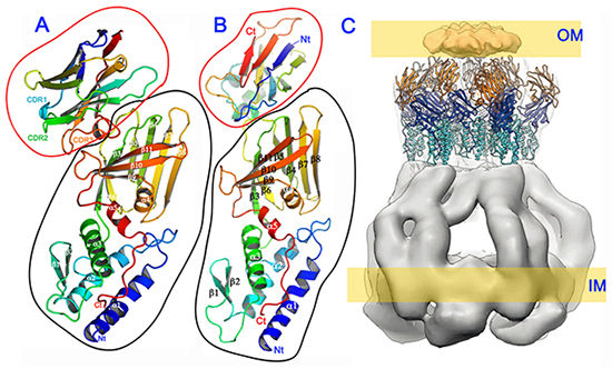
A) Crystal structure of the TssM26Ct–nb25 complex represented as ribbons with TssM26Ct outlined in black and nb25 outlined in red (data collected at SOLEIL-PROXIMA-1)
B) Crystal structure of the TssM26Ct–TssJ complex represented as rainbow-colored ribbons with TssM26Ct outlined in black and TssJ outlined in red (data collected at ESRF ID23-1).
C) Model consisting of 10 copies of the TssM26Ct–TssJ crystal structure docked into the EM volumes corresponding to TssJ and the TssM periplasmic domains 3 and 4 extracted from both the internal and external pillars of the tip complex.