“MAX phases” are materials that combine the properties of metals and ceramics: refractoriness, damage tolerance, resistance to thermal shock, oxidation and corrosion, and conservation of mechanical properties at high temperatures. Thus, they are excellent candidates for applications in extreme environments, like the ones found in reactors. In order to identify the nature of ion irradiation-induced structural damage at the atomic scale, X absorption spectroscopy experiments were carried out on the LUCIA beamline, on irradiated samples of Ti3AlC2.
MAX phases are ternary carbides and nitrides, with chemical formula Mn+1AXn, where M is an early transition metal, A is a group IIIA or IVA element, X is carbon or nitrogen, and n = 1, 2, 3. These materials combine the properties of metals and ceramics, indicating that they could be of interest for use in extreme environments (high temperature, high pressure and radiation). Typically, MAX phases fulfill the requirements for fuel cladding in the core of future fast neutron reactors and gas-cooled fast reactor.
However, identifying the nature of ion irradiation-induced structural damage at the atomic scale is a key to understanding the behavior of MAX phases under irradiation.
First analyses on Ti3AlC2: x-ray diffraction and electron microscopy
The sample was a Ti3AlC2 thin film epitaxially grown by magnetron sputtering onto Al2O3 (0001). Its crystal structure can be described as a stacking sequence of Ti6C octahedra layers interleaved with pure Al element layers. Irradiation experiments were carried out with 240 keV Ar2+ ions at room temperature. The evolution of the structure has been followed by x-ray diffraction (XRD) and transmission electron microscopy (TEM). Although XRD and TEM clearly show evidence of structural damage, they do not provide clear insight into its localization into the structure which remains crystalline but undergoes an ion beam induced phase transformation toward a new phase g. In this context, core-loss spectroscopies are of potential interest for acquiring significant quantitative structural information about the induced damage.
The electron energy loss spectroscopy (EELS) C K edges acquired for the virgin state, irradiated at 5.25 1014 Ar cm-2, and at the highest fluence 1.5 1015 Ar cm-2 remain almost unaffected by the irradiation (figure 1). The C K edge representing essentially the Ti6C octahedra, it is clearly evidenced from EELS that the octahedra layers are extremely stable under ion irradiation up to a dose of 1.7 dpa.
Hence, we can conclude that the stacking of joint octahedra layers is not affected by the ion irradiation. As a consequence, the aluminum layers are likely to be more damaged, accounting for the structural modifications highlighted from XRD and TEM.
The LUCIA x-rays to get more information
To access this information, polarized x-ray absorption spectroscopy (XAS) experiments were performed at SOLEIL on the soft x-ray LUCIA beamline. Two different geometries were used to probe the anisotropy of the charge distribution within the {a, b} planes and along the c axis. In the first one, called in-plane geometry, the sample was set parallel to the x-ray sheet: the incident electric field is then parallel to the sample’s surface and one thus probes the {a, b} planes. In the second one, the out-of-plane geometry, the sample is rotated by 90° with respect to the X-ray beam, the electric field is then along the c axis and we probe this direction. The Al K edges acquired before and after irradiation at the highest fluence are presented in figure 2.
After irradiation, the XANES is strongly modified for both probed geometries, the most salient result being that spectra are nearly undistinguishable. Fine structures are clearly less visible than for the initial state, the edge is smoother, and the first EXAFS energy position is the same, confirming that ion-induced modifications of the Ti3AlC2 crystal structure occur in the aluminum planes. Of particular interest, the XANES after irradiation is, contrary to the initial state, isotropic in shape. Thus the charge distribution around an Al atom is now more isotropic. This can be interpreted as follows: the Al atoms were displaced from a highly anisotropic trigonal prismatic site to a more isotropic atomic site. Another possible assumption is that the Al atoms are organized in a completely random matter, and the Al K edge represents an average of all these positions, and is hence isotropic.
To summarize, from EELS and XAS spectroscopies, the g phase can be viewed as constituted of a ceramic component analogous to the one existing in the parent MAX phase (two joint Ti6C octahedral layers) and a metallic one where the Al atoms seem to be in a cubic symmetry.
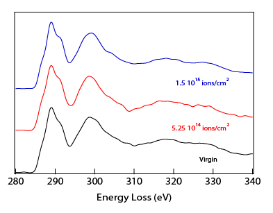
C K EELS for the virgin state (bottom), irradiated at 5.25 1014 Ar cm2 (middle), and at 1.5 1015 Ar cm2 (top)
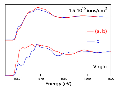
Al K XANES for the virgin state (bottom), and irradiated at 1.5 1015 Ar cm2 (top) for {a, b} and c polarizations.